

Received: 01-Feb-2022, Manuscript No. GJASLPAB-22-59174; Editor assigned: 03-Feb-2022, Pre QC No. GJASLPAB-22-59174 (PQ); Reviewed: 17-Feb-2022, QC No. GJASLPAB-22-59174; Revised: 21-Feb-2022, Manuscript No. GJASLPAB-22-59174 (R); Published: 28-Feb-2022, DOI: 10.35841/ 2408-5499.22.10.174
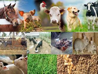
Animal disease is an impairment of the general con- dition of an animal that interferes with or modifies its vital functions. Animal diseases are of major concern because of the economic risks and factors that can cause them to be transmitted to humans. The Depart- ment of Medicine, also known as Veterinary Medicine, deals with the study, prevention, and treatment of diseases not only in pets but also in wildlife and animals used in scientific research. Agriculture related aspects of prevention, control and eradication of economically important animal diseases. Programs related to the control of animal-to-human transmission, especially in pets and wildlife called zoonosis, and are closely linked to human health. Furthermore, the importance of animal diseases is increasing because the primary public-health problem worldwide is animal-protein deficiency in the human diet. However, some animal diseases have been discussed in order to gain a better understanding of the disease.
Foot and Mouth Disease (FMD)
Foot and Mouth Disease (FMD) is a serious, highly contagious disease caused by picarnavirus, a Ribonucleic Acid (RNA) virus of the genus Aptovirus (American Veterinary Medical Association, 2007). There are seven serotypes of FMD virus (O, A, C, SAT (South Africa Territory)1, SAT2, SAT3 and ASIA1). Each serotype of FMD is antigenically different from the other six serotypes. FMD manifests itself in clown oven ruminants such as sheep, cattle, swans, goats, deer and buffaloes (American Veterinary Medical AssociaAssociation, 2007). Humans are not affected by the FMD virus. FMD virus is sensitive to temperature and rapidly inactivates at high temperatures (>56°F). When it comes to survival, the virus has a very narrow window for pH; the optimal range is 7.2-7.6. However, some very reliable data suggest that the virus can be hardy in its natural setting, especially when the conditions are cold and humid.
FMD is especially devastating to livestock. Clinically, FMD is characterized by fever (≥ 41°C) and the development of vesicles (blisters) in the mouth and tongue, lips, nostrils, hooves, and teats of infected animals. Vesicles quickly rupture and form a raw and painful surface. Vesicles cause the animal to salivate profusely and become lame. Infected animals lose weight and produce less milk. Pregnant animals can have abortions. Younger animals may succumb to FMD due to heart damage. Clinical signs vary in severity and the disease is often mild or not evident in sheep. The disease is highly contagious. Inhalation and ingestion of the virus are routes of infection. The virus is primarily transmitted from animal to animal, although other mechanisms such as contaminated feed and fomites are possible. The FMD virus can also be transmitted by contaminated inanimate objects and by people moving between infected and non-infected animals. It is most often spread by transporting infected animals to markets and introducing infected animals to infected flocks. Airborne transmission of FMD virus was recorded; Cattle may be more susceptible to this route of infection. Naturally, the spread of virus cells in the air is dramatically affected by weather conditions.
Avian Influenza
Avian influenza is an infectious disease of birds caused by strains of type A of the influenza virus. A total of 16 hemagglutinin subtypes of type A influenza virus are responsible for the spread of avian influenza. Depending on the severity of the spread and the strain of the virus, avian influenza can be classified as Less Pathogenic Avian Influenza (LPAI) or HPAI. Only hemagglutinin subtypes 5 and 7 cause HPAI. Once introduced, the disease spreads rapidly through domestic poultry herds and wild chicken herds. Because it spreads easily and rapidly from one bird to another, HPAI can have devastating effects, resulting in high mortality in a short period of time. The effects of avian influenza outbreaks are exacerbated by the limited fodder activity found in the poultry industry, the concentrated population of birds and the uniform age of the herd. Avian influenza can occur anywhere in the world. Migratory waterfowl are widely regarded as reservoirs of avian influenza virus. Stool and respiratory secretions contain large amounts of the virus, which can be transmitted to a new host through the conjunctiva or trachea. The avian influenza virus is transmitted by aerosol when birds are in close proximity and by shared drinking water. Infected hens’ eggs may contain the virus, but they are unlikely to survive. Fomites (contact with infected material) and infected birds can spread the disease between flocks. Avian influenza virus can be transmitted through direct contact with infected waterfowl, other infected poultry, surfaces (such as dirt or cages) or contaminated materials (such as water or fodder).
Classical Swine Fever (CSF)
Classical Swine Fever (CSF) is also known as swine fever and hog cholera. The etiologic agent of CSF is a pestivirus, a positive-sense, single-stranded RNA virus of the Flaviviridae family. CSF occurs in both acute and chronic forms. Acute CSF swine is characterized by high fever, inability, and severe depression. In addition, purple skin discoloration, diarrhea, vomiting, cough, weakness of the hind limbs, and eye discharge in severely infected pigs. Pregnant women infected with the CSF virus often have miscarriages. Infected piglets often develop neurological signs, including chills and seizures. There is no cure for CSF. The case-death rate of the acute form of CSF is 95-100% of the suspected population. Death usually occurs within 10-15 days. The CSF virus is transmitted from infected domestic pigs to infected pigs through direct and indirect contact. Taking and inhaling CSF virus is one of the most common routes of infection. The CSF virus can also be transmitted through the transfusion of infected blood or semen. Infected animals excrete the virus in saliva, feces, urine, blood and nasal secretions. Mechanical transmission with contaminated equipment, vehicles, clothing, and footwear (fomites) is possible after the virus has spread. Blood-sucking insects and birds act as mechanical carriers of the CSF virus, although they do not spread it. Consumption of uncooked, infected pork scraps has led to the spread of CSF. The CSF virus can survive for many months in these infected pork products. Transplacental infection with less viral strains produces chronically infected piglets, which can be the source of infection to spread. The incubation period of CSF ranges from 2 to 14 days. After a pig recovers from CSF, it removes the virus for a long time. Thus recovered animals and apparently healthy but infected pigs are important sources of the virus for transmission.