Global Journal of Neurology and Neurosurgery received 111 citations as per Google Scholar report


Received: 01-Jul-2022, Manuscript No. GJNN-22-71845; Editor assigned: 04-Jul-2022, Pre QC No. GJNN-22-71845 (PQ); Reviewed: 18-Jul-2022, QC No. GJNN-22-71845; Revised: 25-Jul-2022, Manuscript No. GJNN-22-71845; Published: 02-Aug-2022, DOI: 10.15651/2449-1942.22.10.006
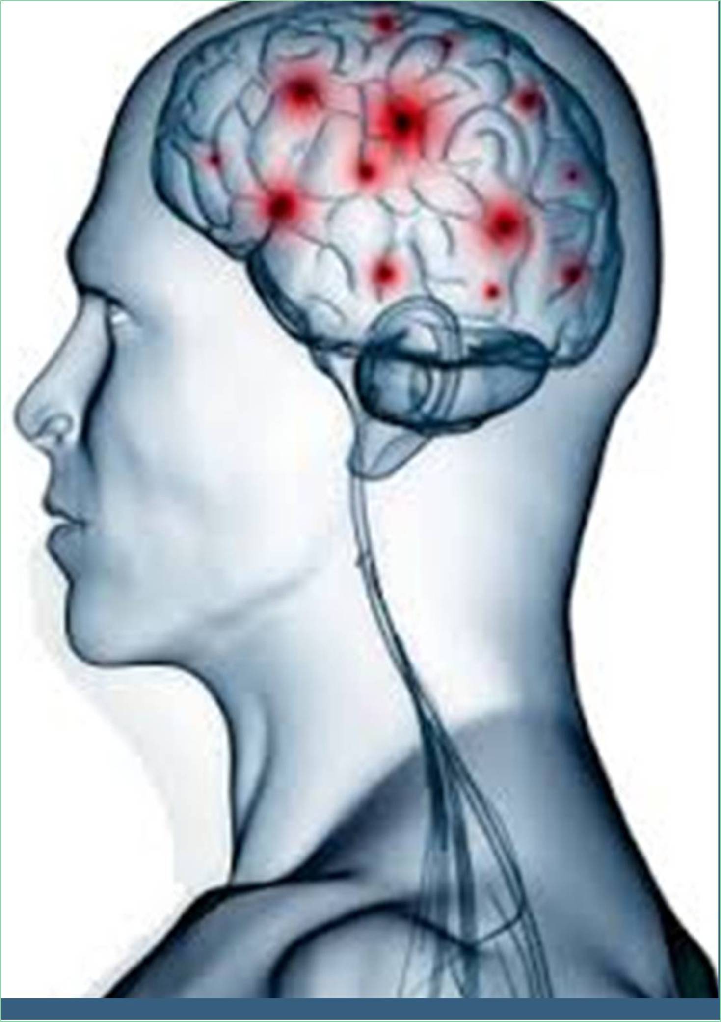
Parts of the nervous system that are not located in the brain and spinal cord are referred to as the peripheral nervous system. The peripheral nerves, neuromuscular junctions, spinal nerves, their roots, and branches are all included. Although the anterior horn cells are technically a component of the Central Nervous System (CNS), because they are a part of the motor unit, they are occasionally discussed with the peripheral nervous system (Zhu, 2020).
Axons, or bundles of nerve fibres, carry information to and from the central nervous system through the peripheral nervous system. The portion of the nervous system responsible for innervating the body's involuntary structures, including the heart, smooth muscles, and glands, is known as the autonomic nervous system. The central and peripheral nervous systems both contain it (Hoffmann, 2018).
Neurones are the name for nerve cells. A neuron is made up of a cell body, which includes a nucleus and cytoplasm, dendrites, which bring electrical impulses into the cell, and an axon, which sends the impulses out of the cell. The dendrites of the neuron after one do not actually touch the axon of that neuron. The synapse is the space between neurons (Chen, 2020).
The complex processes involved in nerve transmission by neurons make up neuronal function. A stimulus, such as light, a specific chemical, or the stretching of a cell membrane by sound, causes the generation of a nerve impulse (action potential) in a sensory neuron. An impulse travels along a neuron from the cell body to the axon through the dendrites (Fehr, 2015).
A chemical transmitter is used to send a signal across a synapse to another neuron. This substance keeps the signal travelling along a nerve by electrically stimulating the subsequent neuron.
Ganglia can be further broken down into sensory ganglia of cranial nerves, spinal nerves (spinal or posterior root ganglia), and autonomic ganglia. Fusiform swellings called sensory ganglia are found on the posterior root of each spinal nerve, close to the root's intersection with the corresponding anterior root. They are also known as posterior root ganglia or spinal ganglia. Sensory ganglia of these nerves are similar ganglia that are also present along the course of cranial nerves V, VII, VIII, IX, and X. The autonomic nervous system's efferent nerve fibres travel along autonomic ganglia, which are frequently asymmetrical in shape. They are located in the paravertebral sympathetic chains, close to or embedded within the walls of various viscera, and around the roots of the great visceral arteries in the abdomen.
The sensory (afferent) division transports sensory signals from central nervous system receptors via afferent Nerve Fibres. It can be divided into somatic and visceral divisions for further subdivision. The viscera of the thoracic and abdominal cavities are the primary sources of signals for the visceral sensory division. The motor (efferent) division sends motor signals from the Central Nervous System (CNS) to effectors via efferent nerve fibres (mainly glands and muscles). It can be divided into somatic and visceral divisions for further subdivision. The skeletal muscles receive signals from the somatic motor division. Signals are sent to glands, cardiac muscle, and smooth muscle by the autonomic nervous system, also known as the visceral motor division. The sympathetic and parasympathetic limbs can be separated from it further (Gao, 2020). The body is typically stimulated to act by the sympathetic division. Generally speaking, the parasympathetic divisions have a calming effect.
Depending on whether they are involved in motor, sensory, somatic, or visceral pathways, PNS nerve fibres are categorised. Motor and sensory fibres can both be found in mixed nerves. The optic and olfactory nerves are examples of sensory nerves, which are less common and mostly comprise sensory fibres. Motor fibres can be found in motor nerves.
Zhu N, Zhang D, Wang W, Li X. (2020). A novel coronavirus from patients with pneumonia in China. New Eng J Med.38:50-78. [Crossref], [Google Scholar], [PubMed]
Hoffmann M, Kleine-Weber H, Schroeder S (2018). SARS-CoV-2 cell entry depends on ACE2 and TMPRSS2 and is blocked by a clinically proven protease inhibitor. Cell. 20:2-20. [Crossref], [Google Scholar], [PubMed]
Chen Y, Liu Q, Guo D (2020). Emerging coronaviruses: genome structure, replication, and pathogenesis. J Med Virol. 92(4): 418-423. [Crossref], [Google Scholar], [PubMed]
Fehr AR, Perlman S (2015). Coronaviruses: an overview of their replication and pathogenesis. Methods Mol Biol.12(82): 1-23. [Crossref], [Google Scholar], [PubMed]
Gao Y, Liming Y, Yucen H, Fengjiang L, Yao Z, Lin C, Tao W (2020). Structure of the RNA-dependent RNA polymerase from COVID-19 virus. Science.64(92):779-782. [Crossref], [Google Scholar], [PubMed]