

Received: 28-Nov-2022, Manuscript No. GJGC-22-83789; Editor assigned: 30-Nov-2022, Pre QC No. GJGC-22-83789 (PQ); Reviewed: 14-Dec-2022, QC No. GJGC-22-83789; Revised: 21-Dec-2022, Manuscript No. GJGC-22-83789 (R); Published: 28-Dec-2022, DOI: 10.15651/GJGC.22.10.015
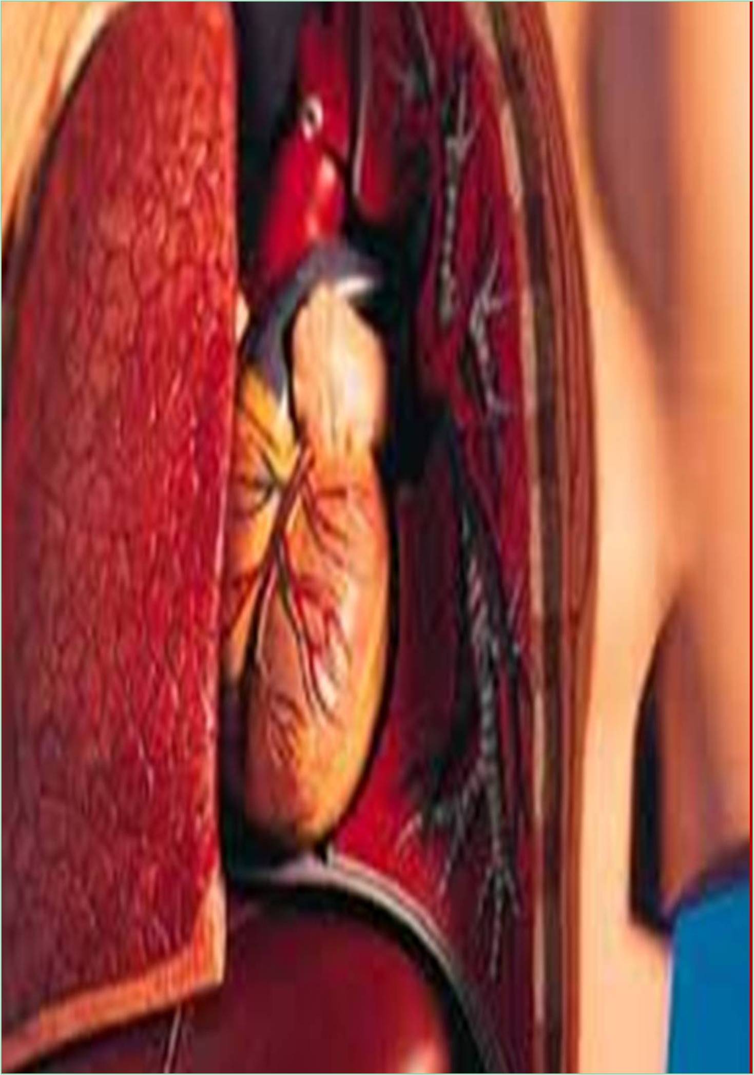
Heart failure is the leading cause of death in developed countries, killing more people than any other disease. It is frequently caused by a scarcity of specific heart muscle cells known as cardiomyocytes, and powerful therapy to repair damaged heart muscle can benefit millions of patients each year. Cardiac remodelling has been thoroughly documented in amphibians, fish, and developing mammals (Grunsfeld et al., 2006). However, following birth, the regeneration of the human heart is confined to the very sluggish replacement of heart muscle cells. Several studies are being conducted to revascularize wounded hearts using adult and pluripotent stem cells, cell reprogramming, and tissue engineering. Although difficult, these therapies may eventually lead to better approaches to treating or preventing heart failure.
The rising prevalence and widespread distribution of heart failure and cardiac disease has fuelled interest in myocardial repair and regeneration studies. The dire and unmet need for new interventional tactics for cardiovascular disease treatment has motivated the rapid deployment of therapeutic procedures to stimulate myocardial regeneration and repair, but the outcomes have been modest thus far (Kile et al., 2006).
Several cellular alterations occur in the myocardium after myocardial infarction. Coagulate necrosis develops over the first 6-12 hours, and fibers near the infarct's perimeter lengthen and narrow, indicating vascular degradation. Edema and neutrophils were found in the intercellular space at the same time. This procedure takes 2-4 days. Following this, necrotic cells are removed by macrophages, which can actively phagocytose for 6 to 10 days. Finally, granulomatous tissue with loose collagen fibers and many capillaries commences the healing and repair process, in which necrotic cardiomyocytes are replaced by a collagen scar (Marin et al., 2005). The immune system is essential for an organism's initial development as well as the continuous replenishment of differentiated cell types to maintain homeostasis. Tissue remodelling, unlike embryonic development, is initiated by trauma, and accumulating evidence suggests that an inflammatory response to this insult also leads regeneration. Many parameters, including age, species, and the accessibility of a population of stem or progenitor cells, influence whether immune activation promotes tissue regeneration or wound healing.
The group investigates the intercellular interactions between cardiac fibroblasts (scar forming cells) and cardiac gonads to understand how these intercellular interventions influence cardiac healing. To examine the topic, they use a mouse model of heart damage and a range of destiny mapping and conditional knockout procedures to change specific genes at various time periods following injury (Starkstein et al., 2008). They went for a signaling pathway of 19 closely related proteins that which play critical roles in organogenesis, wound healing, and cancer treatment.
Proto-oncogene Wnt1 plays a key function in central nervous system development, has recently been shown to serve an important role in controlling the response to fibrous injury in the heart. The heart's fibrous repair response to facilitate regeneration using conditional inactivation and transgenic methods. Embryonic Stem Cells (ESCs) are undifferentiated cells derived from the blastocyst inner cell mass that demonstrate infinite self-renewal and pluripotency (Vijayaraghavan et al., 2002).
They have the ability to grow into endoderm, mesoderm, and ectoderm derivatives. It has been demonstrated that autologous cardiac cell treatments are both safe and efficacious. Understanding the intrinsic mechanisms that regulate endogenous mobilization and/or dispersion of these cells may hold the key to the future of heart repair. These newly discovered endogenous cardiogenic stem cells may be the greatest option for heart repair. These cells should ideally be stimulated in situ, avoiding extraction, purification, culture, and recycling.
Grunsfeld AA, Login IS (2006). Non-alcoholic fatty liver disease and hypertension: coprevalent or correlated? Eur J Gastroenterol Hepatol. 6(1):1-4. [Crossref] [Google Scholar] [PubMed]
Kile SJ, Camilleri CC, Latchaw RE, Tharp BR (2006). Prevalence of liver steatosis and fibrosis detected by transient elastography in adolescents in the 2017-2018 National Health and Nutrition Examination Survey. Clin Gastroenterol Hepatol. 35(6):439-441. [Crossref] [Google Scholar] [PubMed]
Marin RS, Wilkosz PA (2005). Development of new fatty liver, or resolution of existing fatty liver, over five years of follow-up, and risk of incident hypertension. J Hepatol. 20(4):377-388. [Crossref] [Google Scholar] [PubMed]
Starkstein SE, Leentjens AF (2008). Non-alcoholic fatty liver disease: a review of epidemiology, risk factors, diagnosis and management. J Hepatol. 79(10):1088-1092. [Crossref] [Google Scholar] [PubMed]
Vijayaraghavan L, Krishnamoorthy ES, Brown RG, Trimble MR (2002). Clinical association between non-alcoholic fatty liver disease and the development of hypertension. J Gastroenterol Hepatol. 17(5):1052-1057. [Crossref] [Google Scholar] [PubMed]